

Introduction:
Acetabular fractures are challenging orthopaedic injuries because they involve the hip joint and the weight-bearing portion of the pelvis. Displaced and highly multiple pieces acetabular fractures can significantly affect hip stability and joint congruency if not properly reduced and stabilized.
This case demonstrates a complex, multiple pieces acetabular fracture evaluated with three-dimensional CT reconstruction and subsequently treated with open reduction and internal fixation (ORIF) using pelvic/acetabular reconstruction plates and screws.
Case Overview:
The preoperative 3D CT images demonstrate extensive fracture fragmentation involving the acetabular region and adjacent pelvic structures. The fracture pattern is complex, with multiple displaced fragments and disruption of the normal anatomy around the acetabulum.
The postoperative fluoroscopic images demonstrate restoration of the acetabular region with fixation using contoured reconstruction plates and screws.
Patient Profile
- Patient: 61-year-old male
- Injury: Complex acetabular/pelvic fracture
- Imaging: 3D CT reconstruction and intraoperative fluoroscopy
Fracture Characteristics
Displaced, multiple pieces acetabular fracture
Treatment Objectives
Open reduction and internal fixation
Fixation
Pelvic/acetabular reconstruction plates and screws
Preoperative 3D CT Findings & Planning:
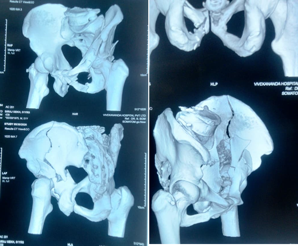
Preoperative Imaging Findings:
The 3D CT reconstruction demonstrates:
- Extensive comminution around the acetabular region
- Multiple displaced osseous fragments
- Involvement of the acetabular columns and surrounding pelvic anatomy
- Disruption of the normal contour of the acetabulum
- Complex fracture extension through the hemipelvis
- Significant displacement requiring anatomical reconstruction
Three-dimensional CT reconstruction is particularly useful in complex acetabular fractures because it allows the surgical team to understand the spatial relationship between the fracture fragments and plan the appropriate surgical approach and fixation strategy.
Fracture Classification
The available images support the diagnosis of a complex acetabular fracture , but an exact Letournel-Judet or AO/OTA subtype cannot be reliably assigned from these photographed 3D reconstructions alone.
For definitive classification, the complete CT dataset should be assessed in axial, coronal and sagittal planes along with the standard AP pelvis and Judet views.
The fracture may represent a complex associated-column acetabular fracture pattern, but the precise subtype should be confirmed by the treating orthopedic surgeon/radiologist.
Surgical Management:
Due to the displacement and comminution, operative stabilization was performed.
The principal objectives of acetabular fracture fixation are:
- Restore the anatomy of the acetabular columns.
- Re-establish a congruent hip joint.
- Restore the weight-bearing surface of the acetabulum.
- Stabilize the fracture fragments.
- Minimize the risk of post-traumatic arthritis.
- Facilitate progressive rehabilitation and functional recovery.
During ORIF, the displaced fracture fragments are mobilized and reduced as anatomically as possible. Reduction clamps, temporary K-wires and appropriately contoured pelvic plates may be used to maintain the reduction before definitive screw fixation.
Implant Specifications:
| Implant | Material | Size / Specification |
|---|---|---|
| Suprapectineal Plate Left 3.5mm | SS316L | 10 Hole |
| Cortex Screw 3.5mm | SS316L | 18mm |
| Cortex Screw 3.5mm | SS316L | 16mm |
| Cortex Screw 3.5mm | SS316L | 15mm |
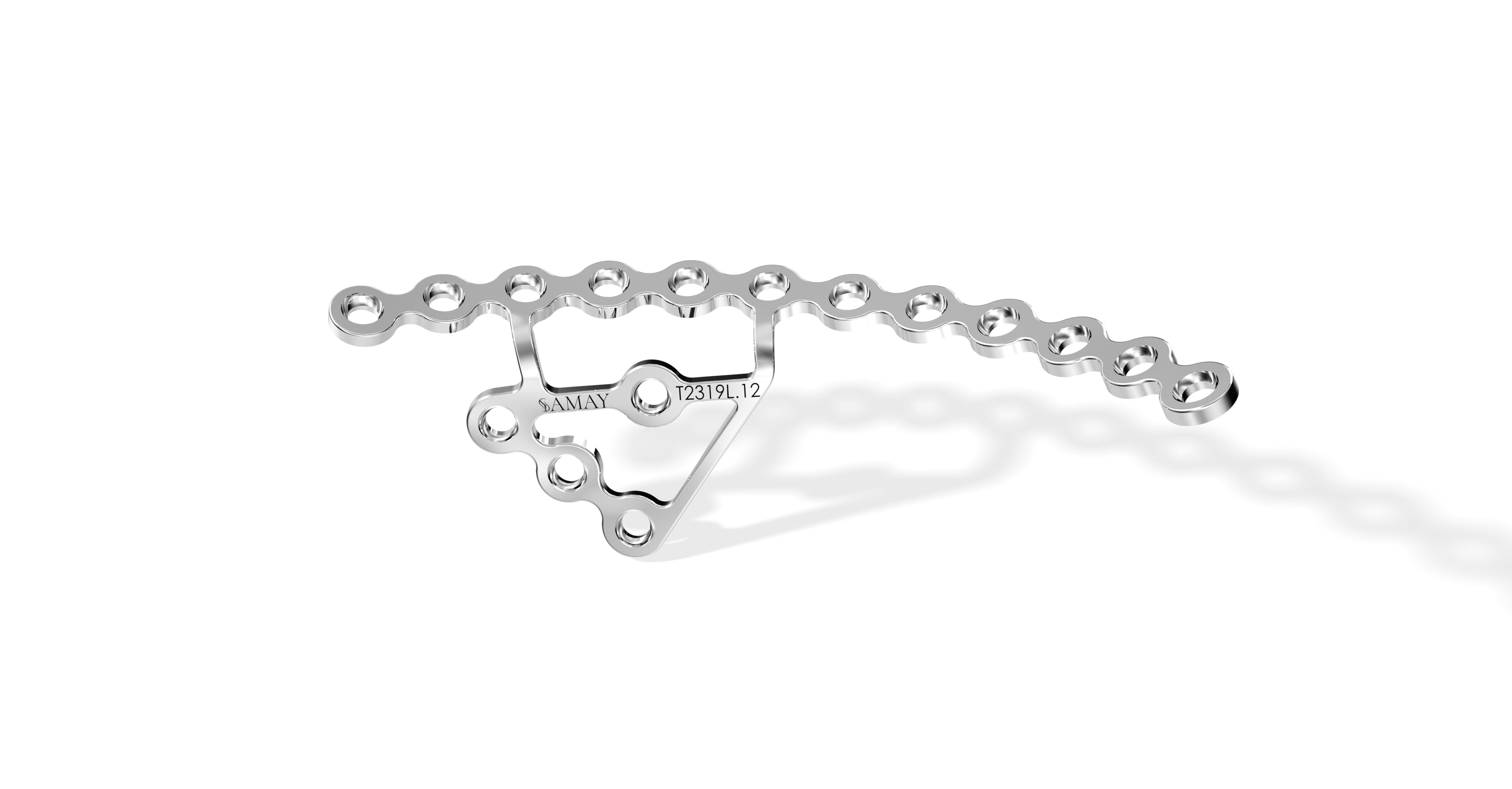
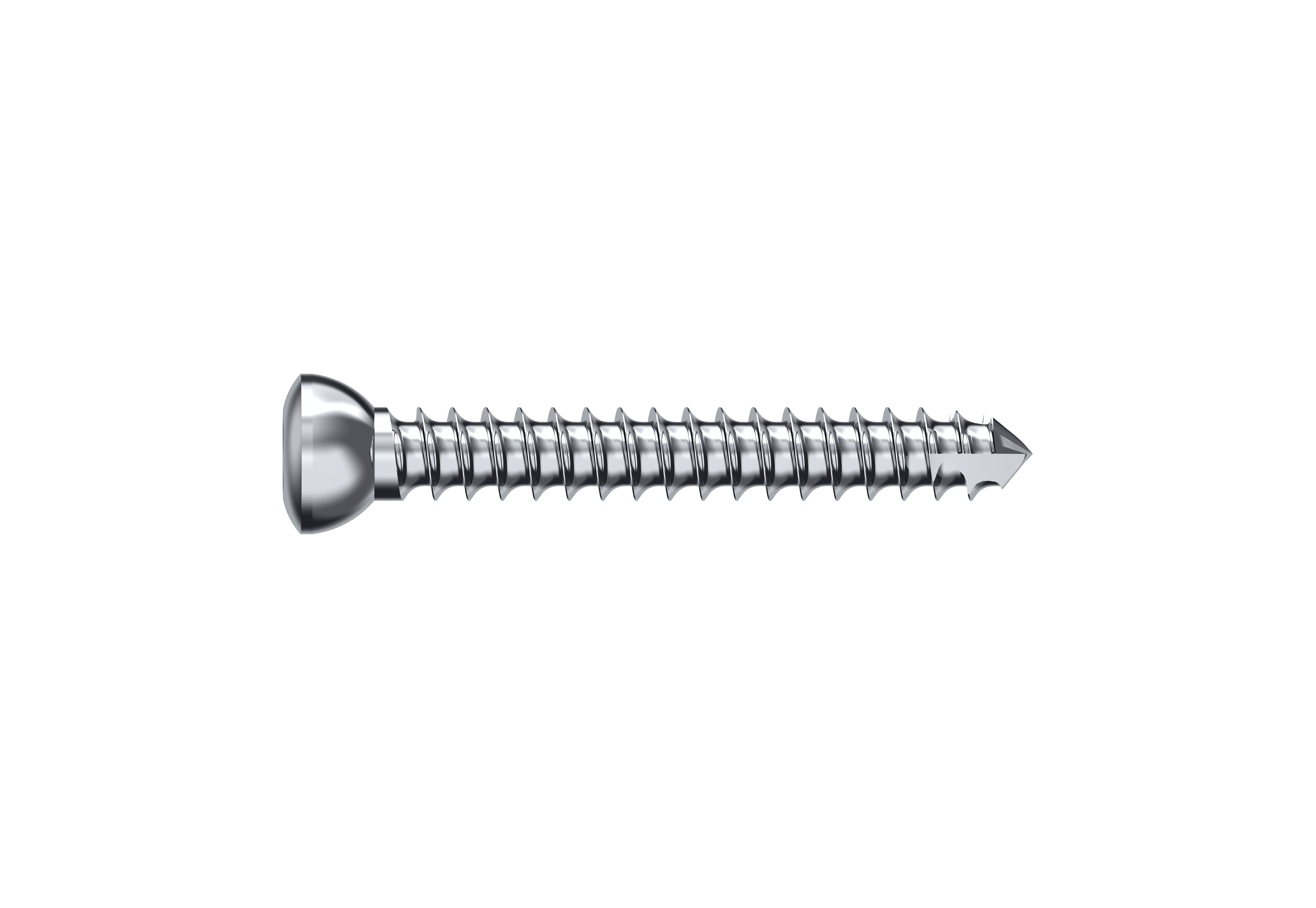
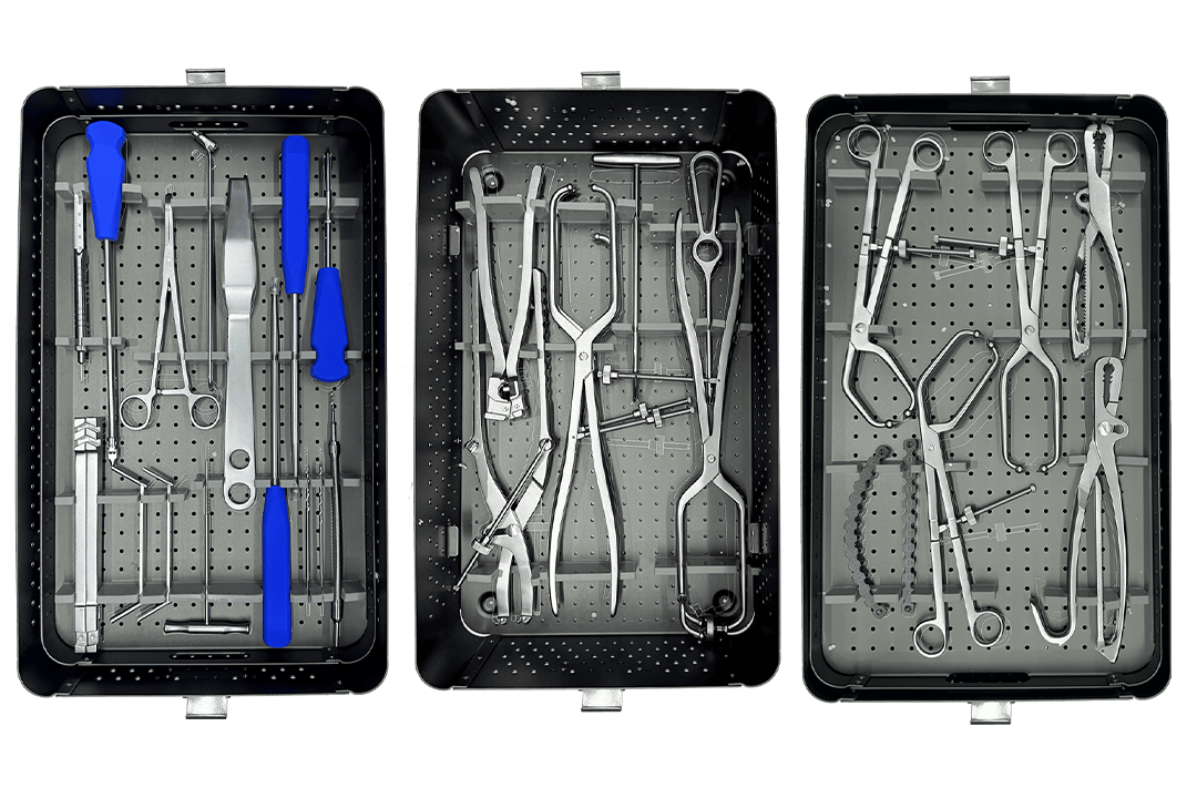
Instrument Set – Pelvic & Acetabular System:
A dedicated pelvic and acetabular instrument set was used for fracture reduction, plate contouring, drilling and screw fixation. The set facilitated accurate reduction and stable fixation of the complex acetabular fracture while supporting efficient intraoperative handling.
Implant Fixation:
The postoperative fluoroscopic images demonstrate fixation with contoured pelvic/acetabular reconstruction plates and screws.
The plates are positioned along the reconstructed acetabular/pelvic columns to provide stable fixation of the fracture fragments.
Appropriate screw placement is particularly important in acetabular surgery because of the proximity of the hip joint and major neurovascular structures.
Postoperative image:
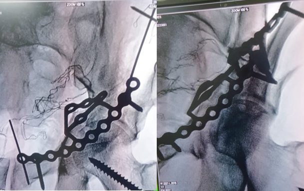
Postoperative Imaging
The postoperative fluoroscopic images demonstrate:
- Improved alignment of the fractured acetabular region
- Stabilization of the major fracture fragments
- Reconstruction plates spanning the involved pelvic region
- Multiple screws providing fixation
- Restoration of the overall acetabular contour
- No obvious gross intra-articular hardware penetration on the provided views
Final assessment of reduction and implant position should be performed using the complete postoperative radiographic series.
Postoperative Care
Postoperative management following complex acetabular fracture fixation generally requires a carefully planned rehabilitation protocol.
Early Postoperative PeriodThe patient is monitored for:
- Neurovascular status
- Wound condition
- Pain and swelling
- Signs of infection
- Deep-vein thrombosis
- Hardware-related complications
Weight-bearing is generally restricted according to fracture stability, bone quality, fixation strength and the surgeon's protocol.
Rehabilitation
A progressive rehabilitation program may include:
- Early ankle and foot exercises
- Quadriceps activation
- Hip and knee range-of-motion exercises as permitted
- Gradual progression of weight-bearing
- Muscle strengthening
- Gait training
The timing of weight-bearing should be individualized by the treating surgeon.
Expected Recovery
Recovery after a complex acetabular fracture can take several months. Radiographic healing, restoration of hip function and the patient's ability to progressively bear weight are important indicators of recovery.
Long-term follow-up is important because acetabular fractures can be associated with complications such as:
- Post-traumatic osteoarthritis
- Heterotopic ossification
- Avascular necrosis of the femoral head
- Sciatic nerve symptoms
- Implant-related complications
- Residual stiffness
- Chronic pain
Importance of Preoperative 3D CT Planning
Complex acetabular fractures can be difficult to understand using plain radiographs alone.
Three-dimensional CT reconstruction provides valuable information regarding:
- Fracture morphology
- Fragment displacement
- Column involvement
- Articular surface involvement
- Surgical approach planning
- Plate positioning
- Screw trajectory planning
For this reason, CT-based evaluation is an important component of treatment planning in complex acetabular trauma.
Conclusion
This case demonstrates the management of a severely displaced and comminuted acetabular fracture requiring operative stabilization.
The preoperative 3D CT images highlight the complexity of the fracture and demonstrate the importance of detailed anatomical assessment before surgery. The postoperative fluoroscopic images show reconstruction and stabilization using contoured acetabular/pelvic reconstruction plates and screws.
Successful management of complex acetabular fractures requires careful preoperative planning, accurate fracture reduction, appropriate implant selection and stable fixation, followed by a structured rehabilitation program.
Frequently Asked Questions:
1. What is an acetabular fracture?
An acetabular fracture is a break involving the socket of the hip joint. Because the acetabulum is part of the weight-bearing hip joint, displaced fractures require careful assessment and, in many cases, surgical fixation.
2. Why is 3D CT used for acetabular fractures?
3D CT helps the surgeon understand the three-dimensional fracture pattern, displacement and involvement of the acetabular columns, facilitating surgical planning.
3. What implants are commonly used for acetabular fracture fixation?
Depending on the fracture pattern, surgeons may use reconstruction plates, pelvic plates, locking plates and appropriately sized screws.
4. Is surgery required for every acetabular fracture?
No. Treatment depends on fracture displacement, hip-joint congruency, stability, fracture pattern, patient factors and associated injuries. Some stable and minimally displaced fractures may be treated conservatively.
5. How long does recovery take after acetabular fracture surgery?
Recovery varies according to fracture severity, fixation stability, bone quality and the patient's overall condition. Rehabilitation and progression of weight-bearing are determined by the treating orthopedic surgeon.
6. Can an acetabular fracture cause arthritis?
Yes. Even after successful fixation, significant acetabular injuries can increase the risk of post-traumatic hip osteoarthritis, particularly when the articular surface has been substantially damaged.